





For nearly three decades, vision scientists believed they understood how the human eye builds its sharpest, most color-rich region. A new discovery from Johns Hopkins University just proved that understanding wrong — and in doing so, opened a promising new path toward future treatments for vision loss.
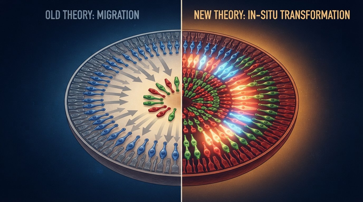
Published in the Proceedings of the National Academy of Sciences, the research reveals that a specialized zone of the retina forms through cellular transformation, not cellular migration — a distinction that fundamentally changes how scientists think about human vision development.
Why The Foveola Matters So Much
At the center of the human retina sits a tiny structure called the foveola — a region so small it makes up only a fraction of the retina’s total area, yet it’s responsible for approximately half of all human visual perception.
Could The Gym Supplement Millions Take For Muscle Also Help Treat Depression?
“This is a key step toward understanding the inner workings of the center of the retina, a critical part of the eye and the first to fail in people with macular degeneration,” said Robert J. Johnston Jr., associate professor of biology at Johns Hopkins and the study’s lead researcher.
Unlike the rest of the retina, which contains all three types of color-sensing cone cells — blue, green, and red — the foveola contains only red and green cones. Understanding exactly how this specialized, blue-free arrangement forms has puzzled vision researchers for decades.
Growing A Retina In A Petri Dish
Studying human retinal development directly has always been extraordinarily difficult, for a simple reason: this process happens before birth, deep inside a developing fetus, making it essentially inaccessible to direct observation.
Compounding the challenge, Johnston notes that common laboratory animals like mice and fish don’t develop the same three-cone-type arrangement found in humans — meaning animal models couldn’t answer this particular question.
To get around this barrier, the Johns Hopkins team used retinal organoids — small clusters of tissue grown in the lab from fetal cells that closely mimic how parts of the human retina actually develop. By observing these lab-grown retinas over several months, researchers were able to directly track the cellular events shaping the foveola as it formed — something never before possible with this level of detail.
The Surprising Discovery: Cells Transform, They Don’t Migrate
The team focused on cone photoreceptors — the light-sensing cells responsible for daytime and color vision, which eventually specialize into blue, green, or red cones, each tuned to a different wavelength of light.
What they observed, tracked over the course of fetal development, was genuinely unexpected.
Between weeks 10 and 12 of development, a small number of blue cones appear in the developing foveola — exactly as the old theory would predict. But by week 14, something remarkable had happened: those same cells were no longer blue cones at all. They had become red and green cones.
The researchers identified two distinct, precisely timed biological mechanisms driving this transformation:
Step one — retinoic acid breakdown: Retinoic acid, a signaling molecule derived directly from vitamin A, is broken down in the developing foveola. This reduces the formation of new blue cones in the region.
Step two — thyroid hormone conversion: Thyroid hormones then act on the remaining blue cones that had already formed, actively converting them into red and green cones.
“First, retinoic acid helps set the pattern. Then, thyroid hormone plays a role in converting the leftover cells,” Johnston explained. “That’s very important because if you have those blue cones in there, you don’t see as well.”
Why This Overturns Decades Of Established Theory
The prevailing scientific model, established roughly 30 years ago, offered a very different explanation for how the foveola achieves its blue-cone-free arrangement.
“The main model in the field from about 30 years ago was that somehow the few blue cones you get in that region just move out of the way,” Johnston said, “that these cells decide what they’re going to be, and they remain this type of cell forever.”
Under this older theory, cone cells were thought to commit permanently to their identity early on — blue cones stayed blue cones, and any blue cones that ended up in the foveola simply relocated elsewhere in the retina, physically clearing space for red and green cones to take their place.
The new evidence tells a fundamentally different story.
“We can’t really rule that out yet, but our data supports a different model,” Johnston said. “These cells actually convert over time, which is really surprising.”
Rather than physically moving, the blue cones appear to remain in place and change their fundamental cellular identity — transforming directly into red and green cones under the precise, sequential influence of vitamin A signaling and thyroid hormone activity.
What This Means For Treating Vision Loss
Beyond resolving a long-standing scientific mystery, this discovery has genuine practical implications for treating diseases that damage vision.
Macular degeneration — a leading cause of vision loss, particularly in older adults, and a disease that currently has no cure — specifically damages the foveola first, the very region this study examined.
Johnston’s team is continuing to refine their retinal organoid models to more closely replicate how the actual human retina functions. Better organoid models could eventually help researchers produce healthier, more accurately specified photoreceptor cells for future cell replacement therapies.
“The goal with using this organoid tech is to eventually make an almost made-to-order population of photoreceptors,” explained Katarzyna Hussey, a co-author of the study now working as a molecular and cell biologist at cell therapy company CiRC Biosciences in Chicago. “A big avenue of potential is cell replacement therapy to introduce healthy cells that can reintegrate into the eye and potentially restore that lost vision.”
Hussey was careful to set realistic expectations about the timeline involved. “These are very long-term experiments, and of course we’d need to do optimizations for safety and efficacy studies prior to moving into the clinic,” she said. “But it’s a viable journey.”
What Comes Next
This research doesn’t yet translate into an available treatment for vision loss. But by identifying the precise biological signals — vitamin A-derived retinoic acid and thyroid hormones — that guide the formation of the human eye’s most important region for sharp vision, scientists now have a clearer roadmap for engineering healthier retinal tissue in the lab.
If future organoid models can reliably reproduce this same cellular transformation process, they could eventually provide a source of specifically differentiated, healthy photoreceptor cells for transplantation — offering new hope for people living with currently untreatable forms of vision loss.
Key Takeaways
- Johns Hopkins researchers discovered that the foveola — the retina’s sharpest vision zone — forms through cellular transformation, not cell migration as previously believed for 30 years
- Blue cone cells convert directly into red and green cones between weeks 10 and 14 of fetal development
- The transformation happens in two stages: vitamin A-derived retinoic acid breakdown, followed by thyroid hormone-driven conversion
- The foveola, though tiny, accounts for roughly half of all human visual perception
- These findings could improve lab-grown retinal organoids and support future cell replacement therapies for macular degeneration
Source: Johns Hopkins University — July 9, 2026
Journal Reference: Katarzyna A. Hussey, Kiara C. Eldred, Brian Guy, et al. A cell fate specification and transition mechanism for human foveolar cone subtype patterning. Proceedings of the National Academy of Sciences, 2026; 123 (7).
DOI: 10.1073/pnas.2510799123