


Multiple sclerosis is one of medicine’s most confounding diseases — not because it’s poorly understood in general, but because it behaves so differently from person to person.
Two individuals can receive the same diagnosis, follow the same treatment protocol, and live completely different lives. One remains relatively stable for decades. The other develops serious disability within years. Why does MS progress rapidly in some people and far more slowly in others?
That question has driven researchers for generations. A new study published in Nature Neuroscience may have found a critical piece of the answer — and it was hiding inside overloaded brain immune cells packed with fat.
Scientists Found That Gen Z Is Biologically Older Than Previous Generations Were At The Same Age
What Multiple Sclerosis Actually Does To The Brain
To understand the discovery, it helps to understand what MS is doing at the cellular level.
MS is an autoimmune disease where the immune system mistakenly attacks myelin — the fatty protective sheath that insulates nerve fibers in the brain and spinal cord. Myelin works like the plastic coating on an electrical wire. When it’s damaged:
- Nerve signals slow down or stop entirely
- Patients develop neurological symptoms including difficulty walking, vision problems, cognitive changes, and fatigue
- The damaged myelin leaves debris that needs to be cleared away for any repair to occur
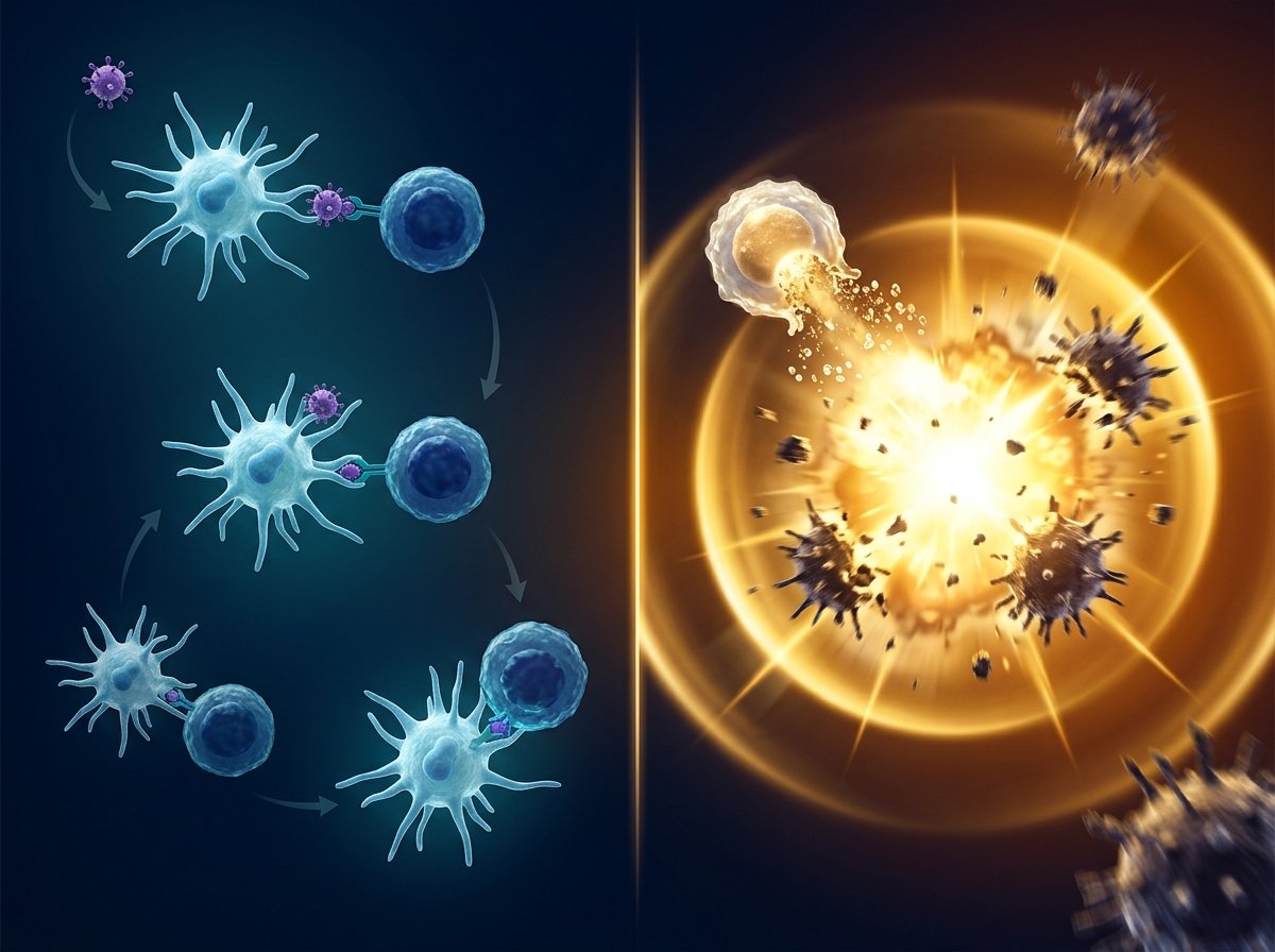
The brain has specialized immune cells designed for exactly this cleanup job: microglia. These cells patrol the brain constantly, removing debris, supporting tissue repair, and maintaining a healthy environment for neurons.
In MS, microglia rush to sites of myelin damage and begin absorbing the debris. And that’s where something goes wrong.
When Cleanup Cells Become Overloaded
Researchers led by Daan van der Vliet at the Netherlands Institute for Neuroscience, working with teams from Leiden University and Utrecht University, examined brain tissue from 28 deceased MS patients who had donated their brains to the Netherlands Brain Bank.
Using a combination of cutting-edge techniques simultaneously — analyzing gene activity, proteins, and fats within individual MS lesions — the team built an unprecedented molecular picture of what was happening in the most severely affected brain regions.
What they found in patients with rapidly progressing, severe MS was striking.
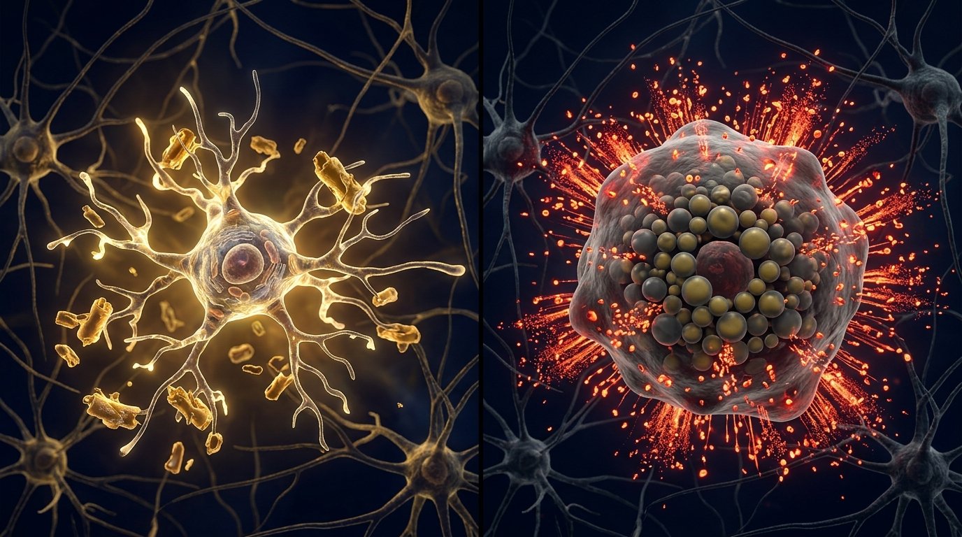
Large numbers of “foamy microglia” — brain immune cells so packed with fat droplets from absorbing damaged myelin that they had taken on a distinctive foamy, bloated appearance — were present throughout the most severely affected areas.
“We found that patients with large numbers of these foamy microglia had a more severe disease course more frequently,” said Van der Vliet.
And more than just being present, these cells appeared to have fundamentally changed what they were doing.
From Brain Protectors To Disease Drivers
Here is the core biological tragedy the study describes.
Microglia absorb damaged myelin as part of their normal cleanup function. In doing so, they take in large amounts of the fats that myelin is composed of. Under normal circumstances, this is manageable — the cells process the material and continue functioning.
But in severe MS, the damage is so extensive that the microglia appear to become overwhelmed. They absorb more fat than they can process. And once overloaded:
- They lose their capacity to repair the surrounding brain tissue
- They become enriched with oxylipins — specific lipid molecules linked to sustained, long-lasting inflammatory activity
- They appear to activate the inflammatory environment rather than resolve it
“These cells are probably trying to do something good: clearing up damage,” Van der Vliet explained. “But they become overloaded, so to speak. As a result, they can no longer effectively contribute to repair.”
The researchers found that MS lesions containing foamy microglia were molecularly distinct from lesions without them — the foamy lesions showed enrichment of specific oxylipin species associated with chronic inflammation, while other lesions did not show this pattern.
A More Complex Picture Of MS Progression
For years, inflammation has been considered the primary driver of MS progression — and current MS treatments largely focus on suppressing the immune system’s attack on myelin.
But this research suggests the story is more complex.
“It does not appear to be simply about the inflammatory response alone,” said Van der Vliet. “These cells are probably attempting to clear damage and promote repair, but that process fails, worsens inflammation, and counteracts recovery.”
In other words, the disease may be partly driving itself through a mechanism that begins as a protective response.
The microglia trying to clean up the damage become overloaded. Overloaded cells release inflammatory fat molecules. Those molecules worsen the inflammatory environment. More myelin is damaged. More microglia become overloaded. And the cycle continues.
This positive feedback loop — where a failed repair mechanism accelerates the very damage it was trying to address — could be a significant contributor to why MS progresses so aggressively in some patients.
The Technology That Made This Possible
The Netherlands Brain Bank was critical to making this research possible.
The ability to study actual human brain tissue — carefully classified and documented over years — gave the research team something that animal models or cell cultures simply cannot provide: a direct window into the biological reality of MS in real human patients, at the molecular level.
“Today we have incredibly sophisticated techniques that can map the brain in great detail,” said Van der Vliet. “The technologies are fantastic, but they tell you relatively little if you cannot connect them to pathology in brain tissue. Precisely because brain tissue has been carefully studied and classified for years by the Netherlands Brain Bank, we were able to recognize these abnormal patterns.”
Toward Future Biomarkers And New Treatments
Perhaps the most clinically significant finding in the study is this: certain oxylipin molecules associated with foamy microglia activity may also be detectable in cerebrospinal fluid — the fluid surrounding the brain and spinal cord that can be sampled through a lumbar puncture.
If confirmed in future studies, these oxylipins could serve as biomarkers — measurable signals that tell doctors, early and objectively, which MS patients are at highest risk of rapid progression and which treatments would be most appropriate for their specific disease biology.
“That opens the possibility of developing biomarkers in the future that could help doctors identify earlier which patients are at risk of rapid decline — and which treatment would suit them best,” said Van der Vliet.
The findings also align directly with current efforts to develop therapies targeting fat metabolism in MS — specifically treatments designed to prevent or reverse the accumulation of damaging lipids in chronic MS lesions. Several experimental treatments in this direction are already being evaluated in clinical studies conducted in collaboration with Roche.
The Bottom Line
MS doesn’t progress the same way in everyone. And now, scientists have a compelling candidate explanation for part of that difference: foamy microglia — brain immune cells overloaded with fat from damaged myelin, switched from repair mode into inflammation mode, fueling the very progression they were meant to prevent.
For the millions of people living with MS worldwide, this discovery doesn’t offer a treatment today. But it offers something equally important: a specific, testable, biologically grounded mechanism that opens new doors for biomarkers, personalized treatment prediction, and a new generation of therapies that target the disease at a level nobody was looking before. 🧠🔬
Source: Netherlands Institute for Neuroscience – KNAW / Nature Neuroscience — June 29, 2026
Journal Reference: Daan van der Vliet, Xinyu Di, Tatiana M. Shamorkina, et al. Foamy microglia link oxylipins to disease progression in multiple sclerosis. Nature Neuroscience, 2026.
DOI: 10.1038/s41593-026-02302-3