









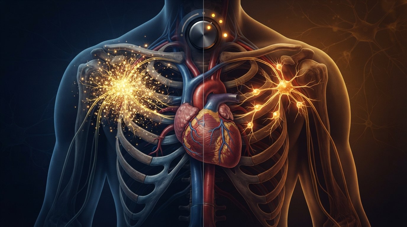
Ask most people what exercise does for the heart, and you’ll get a familiar answer: it makes the heart muscle stronger, improves circulation, and lowers cardiovascular risk over time. All true. But a new study from the University of Bristol reveals something scientists hadn’t fully appreciated before — exercise also physically rewires the nerves that control the heart’s rhythm, and it does so differently on the left and right sides of the body.
Published in Autonomic Neuroscience, the research could open the door to more precise, personalized treatments for common heart conditions including arrhythmias, angina, and stress-induced “broken-heart” syndrome.
Meet The Heart’s “Dimmer Switch”
To understand this discovery, it helps to understand a specific and often-overlooked part of the nervous system: the stellate ganglia.
Nanoplastics In Drinking Water May Be Making Dangerous Bacteria Stronger
These are paired clusters of nerve cells located in the lower neck and upper chest, on both the left and right sides of the body. Their job is to send “go faster” signals to the heart, forming a critical part of the sympathetic nervous system — the branch of the nervous system responsible for the body’s rapid response to stress, exertion, and physical demand.
“These nerve clusters act like the heart’s dimmer switch,” explained study lead author Dr. Augusto Coppi, Senior Lecturer in Veterinary Anatomy at the University of Bristol.
Just as a dimmer switch allows fine control over a light’s brightness rather than a simple on/off, the stellate ganglia allow fine control over how fast and forcefully the heart beats — adjusting continuously based on physical demand, stress, and other bodily signals.
An International Research Collaboration
The study was carried out through collaboration between the University of Bristol, University College London (UCL) in the UK, the University of São Paulo (USP), and the Federal University of São Paulo (UNIFESP) in Brazil.
Using advanced three-dimensional imaging techniques known as stereology — a precise method for quantifying microscopic structures in tissue — the research team examined exactly how aerobic exercise changed the nerve clusters responsible for regulating heart function.
What The Researchers Found
After 10 weeks of aerobic training, the exercised rats showed dramatic and distinctly side-specific changes in their stellate ganglia compared to untrained animals.
On the right side, the cardiovascular nerve cluster contained roughly four times as many neurons as in untrained animals — a substantial increase in the sheer number of nerve cells present.
On the left side, something different happened: rather than increasing in number, the existing neurons nearly doubled in size. Meanwhile, on the right side, individual neurons actually became slightly smaller, even as their overall numbers increased dramatically.
This is a genuinely striking pattern. Exercise wasn’t simply “strengthening” the heart’s nerve supply in a generic, uniform way — it was reshaping the left and right nerve clusters through two entirely different structural strategies: more numerous but smaller neurons on the right, fewer but larger neurons on the left.
“The discovery points to a previously hidden left-right pattern in the body’s ‘autopilot’ system that helps run the heart,” Coppi said. “We’ve shown that regular, moderate exercise remodels that switch in a side-specific way. This could help explain why some treatments work better on one side than the other and, in future, help doctors target therapies more precisely and effectively.”
Why This Matters For Treating Heart Conditions
This discovery has direct relevance to several serious and common heart conditions that are already treated by targeting the stellate ganglia.
“Irregular heart rhythms, known as arrhythmias, stress-induced ‘broken-heart’ syndrome, and certain types of chest pain are often treated by dialing down overactive stellate ganglia,” Coppi explained.
Current treatment approaches for these conditions sometimes involve:
- Nerve blocks — temporarily interrupting nerve signals from the stellate ganglia to reduce overactive cardiac signaling
- Denervation procedures — more permanent interventions that reduce nerve activity in these clusters to help manage severe or treatment-resistant arrhythmias
Currently, these procedures are often performed without detailed consideration of how the left and right stellate ganglia might genuinely differ from one another — either naturally, or as a result of a patient’s activity levels, fitness, and lifestyle.
“By mapping how exercise changes these ganglia on each side, the study offers clues that could one day fine-tune procedures like nerve blocks or denervation to the side most likely to help,” Coppi said.
In practical terms: if doctors eventually understand which side of a patient’s stellate ganglia is driving a particular rhythm problem — and how that side’s structure has been shaped by factors like exercise history — treatment could become significantly more targeted and personalized, rather than relying on more generalized approaches.
Important Limitations To Understand
Coppi is direct about where this research currently stands. “The findings are early-stage and in rats, so clinical studies would need to follow,” he said.
This distinction matters. While rat models of cardiovascular physiology often translate meaningfully to human biology, confirmation in humans is essential before any of these findings could influence actual clinical practice. The structural and functional relevance of these left-right differences in human hearts remains to be established.
What Comes Next
The research team has clearly defined next steps for building on these initial findings.
They plan to investigate how these structural nerve changes actually affect heart performance, both during exercise and at rest — moving from anatomical observation toward functional understanding of what these rewired nerve clusters actually do to heart rhythm and response.
They also intend to determine whether the same left-right asymmetric pattern appears in other animal models, and critically, whether it can be detected in humans using non-invasive markers — a necessary step before any of this research could meaningfully inform human cardiac treatment.
“Understanding these left-right differences could help us personalize treatments for heart rhythm disorders and angina,” Coppi said. “Our next step is to test how these structural changes map onto function and whether similar patterns appear in larger animals and humans.”
Why This Represents A Meaningful Shift In Cardiology
This research reflects a broader and important direction in modern medicine: moving away from generalized, one-size-fits-all treatment approaches and toward genuinely personalized medicine — treatments informed by an individual’s specific physiology, structure, and biological history.
If confirmed in humans, understanding side-specific nerve remodeling from exercise could eventually help clinicians make more informed decisions about which side of a patient’s nervous system to target during interventions for arrhythmias, angina, or stress-related cardiac events — turning what has traditionally been a somewhat generalized procedure into a more precisely tailored one.
For now, the discovery adds a fascinating new dimension to something we already knew was good for us. Exercise strengthens the heart. It also, it turns out, quietly rewires the nervous system that keeps it beating — in ways scientists are only just beginning to map. 🫀🧬
Key Takeaways
- Exercise physically remodels the stellate ganglia — paired nerve clusters that regulate heart rhythm — differently on the left and right sides of the body
- After 10 weeks of aerobic training, right-side nerve clusters showed roughly 4 times more neurons, while left-side neurons nearly doubled in size
- This left-right asymmetry could help explain why some heart treatments, like nerve blocks or denervation, work better on one side than the other
- The findings could eventually help doctors develop more precise, personalized treatments for arrhythmias, angina, and stress-induced heart conditions
- This is early-stage research conducted in rats; human studies are needed to confirm these findings
Source: University of Bristol — July 13, 2026
Journal Reference: Fernando Vagner Lobo Ladd, Aliny Antunes Barbosa, Renato Albuquerque de Oliveira Cavalcanti, et al. Asymmetric neuroplasticity in stellate ganglia: Unveiling side-specific adaptations to aerobic exercise. Autonomic Neuroscience, 2025; 262: 103338.
DOI: 10.1016/j.autneu.2025.103338