







Alzheimer’s disease affects more than 7 million Americans, and while there is currently no cure, doctors have long relied on selective serotonin reuptake inhibitors (SSRIs) to help manage the neuropsychiatric symptoms — anxiety, depression, and agitation — that affect nearly all patients at some stage of the disease.
The problem is that responses to these medications vary enormously from person to person, and until now, there’s been no reliable way to predict who will actually benefit. Scientists at Johns Hopkins Medicine may have just found a solution — grown, quite literally, from patients’ own blood.
Published in Alzheimer’s & Dementia, the study demonstrates that miniature, lab-grown brain tissue models called organoids could help predict individual drug response and eventually guide truly personalized Alzheimer’s treatment.
Turning Blood Into Brain Tissue
The research began with blood samples collected, with permission, from patients enrolled at the NIH-funded Johns Hopkins Alzheimer’s Disease Research Center.
Exercise Doesn’t Just Strengthen The Heart. It Rewires It
Using a technique that has transformed regenerative medicine over the past two decades, researchers reprogrammed these blood cells back into a stem cell-like state — creating what are known as induced pluripotent stem cells (iPSCs). These remarkable cells retain the ability to develop into virtually any cell type in the human body.
From these patient-derived stem cells, the team guided the development of hindbrain organoids — small, pea-sized clusters of brain tissue that closely resemble the hindbrain, a region at the base of the skull responsible for essential functions like breathing, sleep, and heart rate regulation. Importantly, these organoids contained specialized neurons capable of producing serotonin — the neurotransmitter that SSRIs specifically target.
The scale of this effort was genuinely significant. The study included hundreds of organoids representing individual Alzheimer’s patients alongside organoids grown from healthy participants — making it, according to lead researcher Dr. Vasiliki Machairaki, potentially one of the largest brain organoid studies ever conducted in Alzheimer’s research.
Mini Brains That Actually Reproduce Alzheimer’s Biology
A critical first question was whether these lab-grown organoids would genuinely reflect real Alzheimer’s-related biology — or simply behave like generic brain tissue regardless of the donor’s disease status.
The answer was clear. Compared with organoids grown from healthy individuals, those derived from Alzheimer’s patients showed distinct differences in proteins involved in communication between brain cells, inflammation, and pathways specifically associated with the disease.
This validation step matters enormously. It confirms that these patient-specific organoids weren’t just generic brain tissue grown in a dish — they were capturing something biologically meaningful and disease-relevant about each individual patient’s underlying condition.
Testing A Common Antidepressant On Patient-Specific Tissue
With disease-relevant organoids established, the research team moved to the study’s central question: could these mini brains reveal how different patients’ tissue might respond to escitalopram oxalate, a widely prescribed SSRI antidepressant?
The results revealed striking variation between patients.
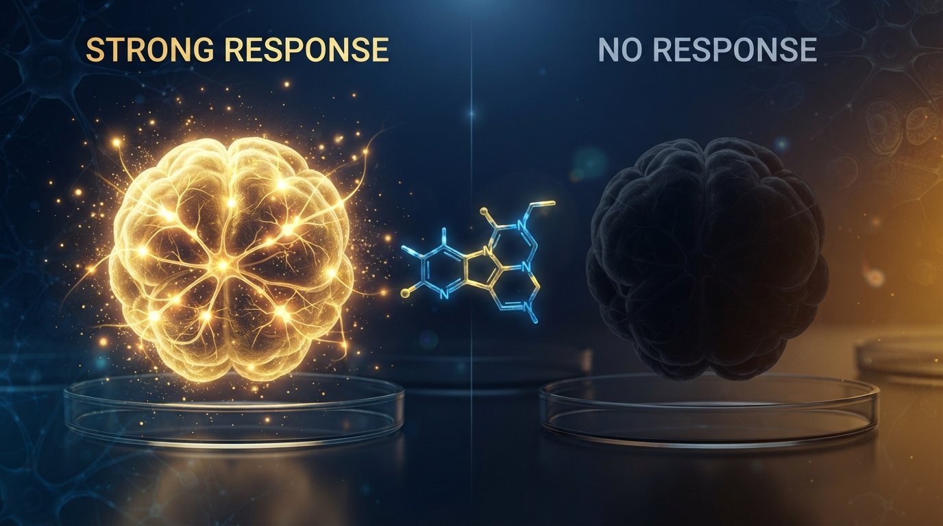
In some patient-derived organoids, escitalopram treatment increased proteins involved in serotonin signaling and communication between brain cells — exactly the biological pathways antidepressants are designed to influence. This indicated the tissue was genuinely responding to the medication at a molecular level.
Other organoids, derived from different patients, showed little to no molecular response to the same treatment at the same dose.
“We used these organoids to model how some patients’ tissue may respond to a commonly prescribed SSRI,” Machairaki explained. “On a large-scale level, our model may eventually be used to identify subgroups of patients, based on underlying molecular mechanisms, who are more likely to respond to certain drugs and thus help us to create precise, targeted treatments in the long run.”
This is the essence of precision medicine applied directly to Alzheimer’s care — rather than prescribing the same medication to every patient and waiting weeks or months to see if it works, doctors could eventually test a patient’s own organoid model first, predicting response before treatment even begins.
Tiny Particles That May Reveal Disease Stage And Drug Response
Beyond the organoids themselves, the research team made a second significant discovery involving extracellular vesicles — tiny particles that cells release, carrying proteins and other molecular cargo that reflect what’s happening inside the cell of origin.
Before and after treating the organoids with escitalopram, researchers examined the proteins contained within these vesicles, comparing patient-derived and healthy control organoids.
The vesicles contained proteins involved in essential brain functions — communication between neurons, memory formation, and neurotransmitter release. Alzheimer’s-derived organoids showed clear reductions in several specific disease-associated proteins, including RAB3A, NSF, and ATCAY — all of which play important roles in normal signaling between brain cells.
After escitalopram treatment, protein levels increased in certain samples, particularly proteins connected to serotonin signaling and synaptic pathways — mirroring the variation seen directly in the organoid tissue itself. Some samples showed strong molecular responses; others showed little to none.
“This variation raises the possibility that extracellular vesicles from brain organoids could eventually help identify which patients are most likely to benefit from a particular treatment,” according to the research team’s findings.
Toward A Future “Liquid Biopsy” For Alzheimer’s
Perhaps the most exciting long-term implication of this research involves these extracellular vesicles becoming a genuine diagnostic tool.
Because these particles can potentially be detected and analyzed from blood samples — a far less invasive approach than brain imaging or spinal fluid analysis — Machairaki hopes they might eventually function as a kind of liquid biopsy for Alzheimer’s disease: a relatively simple blood test capable of helping diagnose the condition, determine its stage of progression, and identify a patient’s specific disease subtype.
Machairaki is careful to frame this as an early step rather than an imminent clinical tool. “Our study suggests that large-scale, patient-derived brain organoids and the vesicles they secrete can help us stage Alzheimer’s disease, investigate the mechanisms that drive it and assess how patient subgroups may respond to different treatments,” she said.
Building More Realistic Brain Models
Looking ahead, Machairaki plans to develop more advanced organoids that incorporate additional biological complexity — specifically, immune cells and vascular-like networks that mimic real blood vessels.
Adding these features would bring the lab-grown tissue significantly closer to resembling actual living human brain tissue, where immune activity and blood supply play crucial roles in both healthy function and disease progression — factors the current organoid models don’t yet fully capture.
Why This Research Matters
Alzheimer’s disease has long been treated with a relatively uniform approach to managing associated psychiatric symptoms, despite clear clinical evidence that patients respond very differently to the same medications. This has meant a frustrating, often lengthy process of trial and error for patients and caregivers alike — trying one medication, waiting to see if it helps, then trying another if it doesn’t.
If patient-derived brain organoids can reliably predict drug response before treatment begins, it could meaningfully shorten that difficult trial-and-error period, sparing patients and families from weeks or months of ineffective treatment while pointing directly toward the medications most likely to actually help.
Key Takeaways
- Johns Hopkins researchers grew hundreds of pea-sized “hindbrain organoids” from the reprogrammed blood cells of Alzheimer’s patients
- These patient-derived organoids reproduced real molecular features of Alzheimer’s disease, including altered inflammation and cell-communication proteins
- When treated with the SSRI escitalopram, some organoids showed strong molecular responses while others showed almost none — mirroring the variable drug response seen in real patients
- The organoids released extracellular vesicles containing disease-related proteins, which could eventually serve as blood-based biomarkers for diagnosing and staging Alzheimer’s
- This research represents an early but promising step toward personalized, precision-based Alzheimer’s treatment
Source: Johns Hopkins Medicine — July 22, 2026
Journal Reference: Rachel J. Boyd, Daiyun Dong, Ram Sagar, et al. Proteomic profiling of brain organoids and extracellular vesicles identifies early Alzheimer’s disease biomarkers and drug response heterogeneity. Alzheimer’s & Dementia, 2026; 22 (4).
DOI: 10.1002/alz.71273